| 0 | |
| Sara Abrahamsson Multifocus SIM and the challenge of live 3D super-resolution imaging University of California, Santa Cruz | |
| Hillel Adesnik Three dimensional holographic optogenetics University of California, Berkeley | |
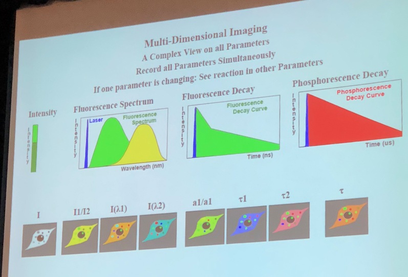
| Wolfgang Becker The Multi-Dimensional World of TCSPC FLIM Becker-Hickl, GmbH | |
| Wolfgang Becker II Fast-Acquisition TCSPC FLIM with sub-25 ps IRF Width Becker-Hickl | |
| Eric Betzig Design of a multimodal “Swiss army knife” microscope for in vivo adaptive optical imaging of diverse specimens University of California, Berkeley, and Janelia Research Campus, HHMI | |
| Martin Booth Advances in Dynamic Optics for Microscopy University of Oxford | |
| Lukas Braun Assessment of clinically relevant metabolic parameters by TCSPC fluorescence lifetime imaging: A new approach with 1p-excitation on the DCS-120 Confocal FLIM System Becker & Hickl GmbH, Berlin, Germany | |
| Teng-Leong Chew Janelia Advanced Imaging Center: Challenges and Impacts from Open Access on a Global Scale HHMI Janelia Research Campus | |
| Wonshik Choi Simultaneous suppression of aberration and multiple scattering in optical coherence imaging Korea University | |
| Beth Cimini Getting the most from your images with morphological profiling Broad Institute | |
| Carlo Condello Fluorescent amyloid multiple emission spectra (FLAMES) microscopy to decipher structural variants of misfolded proteins University of California, San Francisco | |
| Sheel Dodani Genetically Encoded Fluorescent Sensors to Illuminate Cellular Chloride Signaling University of Texas at Dallas | |
| Alfredo Dubra Shack-Hartmann centroiding: back to basics Stanford University | |
| Kevin Eliceiri The ImageJ Ecosystem: An Open and Extensible Platform for Biomedical Image Analysis University of Wisconsin, Madison | |
| Samantha Fore ZEISS Elyra 7 with Lattice SIM, a new Platform for Fast and Gentle 3D Superresolution Microscopy Zeiss Microscopy | |
| Ruixuan Gao Scalable imaging of brain ultrastructure with molecular contrast and nanoscale resolution MIT and Janelia Research Campus, HHMI | |
| Galo Garcia A super-resolution view of signaling through the cell's antenna University of California, San Francisco | |
| Thomas Gensch Flavin-binding fluorescent proteins as genetically encoded photosensitizers Forschungszentrum Juelich | |
| Elizabeth Hillman SCAPE microscopy for even faster in-vivo 3D microscopy Columbia University | |
| Na Ji High-resolution high-speed recording of neural activity University of California, Berkeley | |
| 0 | Karsten König Multiphoton-FLIM-Tomography of Solar Cells JenLab |
| Julia Lazzari-Dean Optical Quantification of Outer Membrane Voltage in Growth Factor Signaling University of California, Berkeley | |
| Yolanda Markaki Quantitative 3D-SIM imaging of chromatin organisation; Insights into X-chromosome inactivation and formation of repressive nuclear compartments University of California, Los Angeles | |
| 0 | Sabrina Matthias Improving quantitative fluorescence imaging with flat field illumination Asphericon |
| Dane Maxfield Cellular Imaging with Light-Sheet Fluorescence Microscopy: Ultra Gentle, High-Resolution Imaging of Living Samples Bruker | |
| Cynthia McMurray Spectral phenotyping by infrared light Lawrence Berkeley National Laboratory | |
| Sohum Mehta Fluorescent biosensors for multiplexed imaging of signaling activities in living cells. University of California, San Diego | |
| Yves Mely Advanced imaging techniques to study the interactions and dynamics of viral and bacterial proteins University of Strasbourg | |
| Zachary Newman Nanoscale structure-function analysis of synaptic transmission at the Drosophila larval neuromuscular junction University of California, Berkeley | |
| Medha Pathak Piezo1 activation gains traction University of California, Irvine | |
| Ammasi Periasamy Metabolic Mapping of Cancer cells and tissues - Multiphoton FLIM-FRET Microscopy University of Virginia | |
| Hesper Rego Imaging a bacterial infection, one cell at a time Yale University | |
| Austin Roorda Testing human vision in a cellular scale University of California, Berkeley | |
| Balázs Rózsa Fast 3D imaging and re-activation of neuronal networks, dendrites, and spines in several cubic millimeter volumes in behaving animals to understand visual representation Hungarian Academy of Sciences | |
| Angelika Rück Basics and applications of metabolic FLIM and oxygen PLIM University of Ulm | |
| 0 | Lydia Sauer Shifted-Component Model Improves Ophthalmic FLIM Data Analysis University of Utah |
| Johannes Schöneberg Advanced methods in microscopy and solutions to the resulting big data challenges University of California, Berkeley | |
| Ben Shababo High-throughput, cellular-resolution neural circuit mapping with two-photon optogenetics and computational experimental design University of California, Berkeley | |
| 0 | Matthew Shaw Superconducting Nanowire Single Photon Detectors Jet Propulsion Laboratory, NASA |
| Vladislav Shcheslavskiy Time-resolved macroimaging and spectroscopy Becker-Hickl | |
| Melissa Skala Label-free Classification of T cell Activation University of Wisconsin, Madison | |
| Andrea Slade Correlative Light Microscopy and AFM for Live Cell and Tissue Studies Bruker | |
| Ellen Sletten Flavylium polymethine fluorophores for imaging in the shortwave infrared University of California, Los Angeles | |
| Klaus Suhling Fluorescence Lifetime Imaging (FLIM) for viscosity and diffusion measurements King's College London | |
| Srigokul Upadhyayula Imaging Subcellular Dynamics in Multicellular Organisms Harvard Medical School | |
| Steven Vogel Venus(A206) dimers behave coherently at room temperature National Institutes of Health | |
| Winfried Wiegraebe Mapping A Human Stem Cell State Space Using A Microscopy Pipeline Allen Institute for Cell Science | |
| Graham Wright Probing the structure and function of the nucleus with 3D-SIM: Applications studying laminopathies and ageing Skin Research Institute of Singapore, A*STAR | |
| Ke Xu Multifunctional & multidimensional super-resolution microscopy University of California, Berkeley | |
| Andrew York Photobleaching and Stimulated Emission: Two Stories About New Imaging Tricks Using Old Photophysics Calico Labs | |
| 0 | Elena Zagaynova Multimodal optical imaging: MPT, FLIM, OCT MA, for label free clinical diagnosis Institute of Biomedical Technologies |
| Maxim Ziatdinov Deep machine learning for atom-resolved imaging Oak Ridge National Laboratory | |